QP-Insights®
The value of medical imaging is hard to unlock
Imaging data is often fragmented across systems, acquired in different ways, and difficult to standardize and quantify. Reads are manual and vary between readers. Evidence gets assembled by hand — endpoints stitched from spreadsheets in research, quantification done manually if at all in the clinic — and there is no reliable way to trace any result back to the underlying data.
Furthermore, exploratory questions mostly go unasked because the data was never structured for analysis and the models to explore it weren’t within reach.
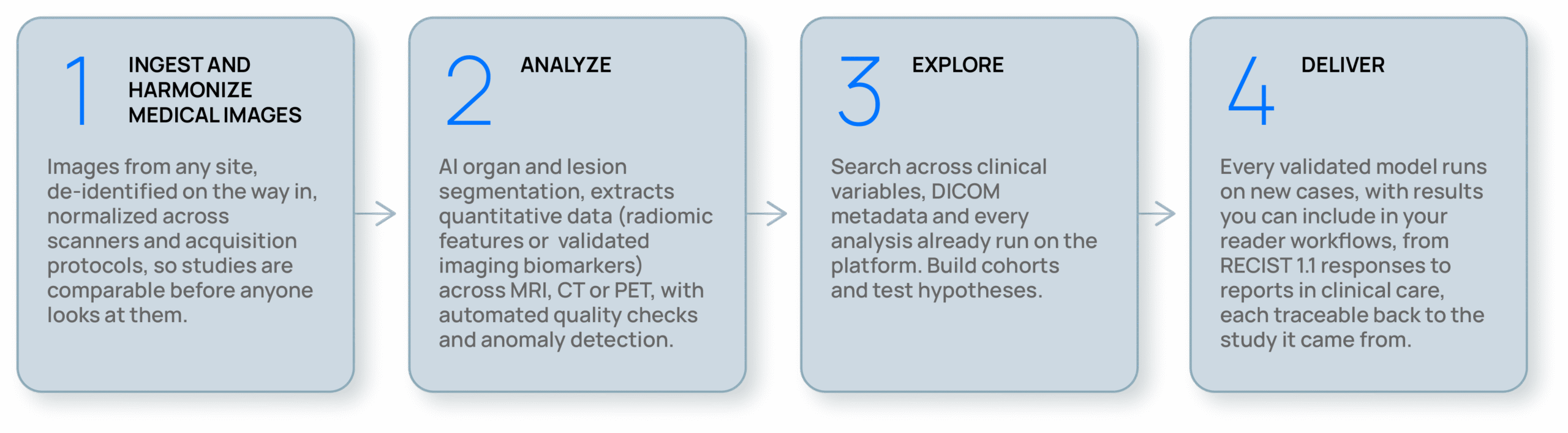
QP-Insights® replaces all that with one system. Data is harmonized on the way in, analyzed consistently, queryable at any point, and auditable from the endpoint back to the voxel.
QP-Insights® is Quibim’s multimodal imaging AI platform that transforms imaging, together with molecular and clinical data, into rigorously validated models and provides the environment where those models run seamlessly, from early research all the way into clinical care.
How it works
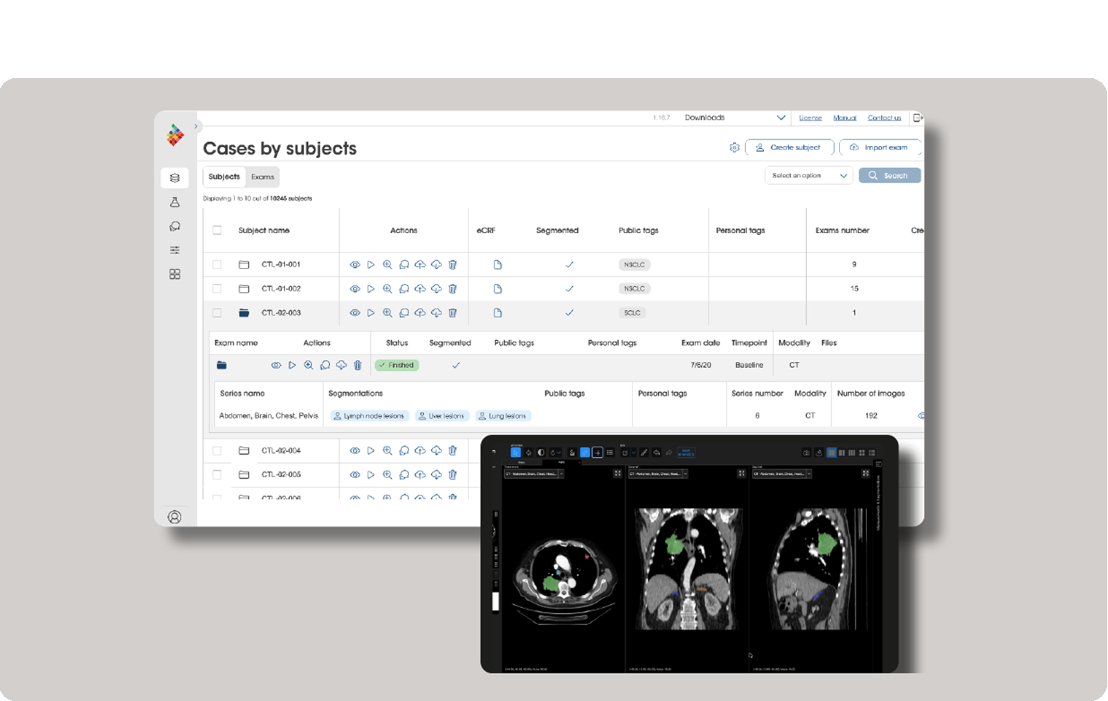
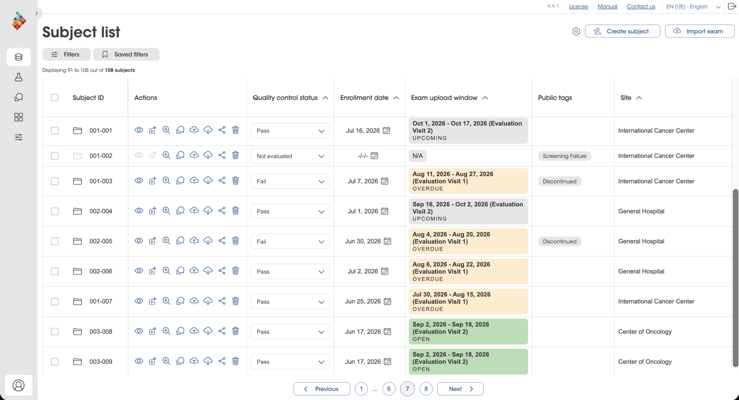
A multi-omics data management platform
Configurable to how your program runs.
Zero-footprint viewer
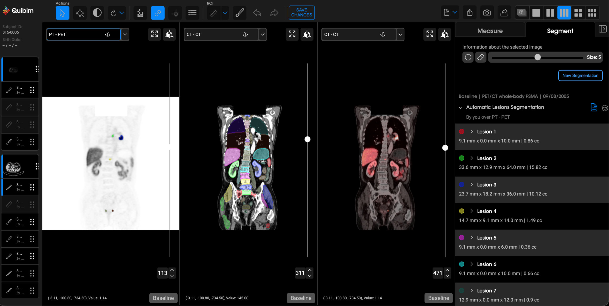
Load, display and annotate DICOM images directly in the browser with no installations or local setup required. Users can mark findings and regions of interest, and segment organs using our integrated tools. The same viewer also powers our regulatory-cleared products, providing compliant image visualization when used in clinical workflows.

AI-driven organ & lesion segmentation
An organ-agnostic methodology that operates across modalities, anatomies and conditions — paired with automated quality checks and anomaly detection so failures surface immediately rather than silently propagating.

Imaging biomarkers & radiomics
Reveal what is already embedded in every conventional scan: quantitative signal that strengthens diagnostic accuracy, prognosis and prediction of treatment response.
Model deployment
Once a model is developed and validated, it can be seamlessly integrated into QP-Insights® enabling immediate impact on new cases and delivering results across clinical trials and routine patient care.
Cohort builder
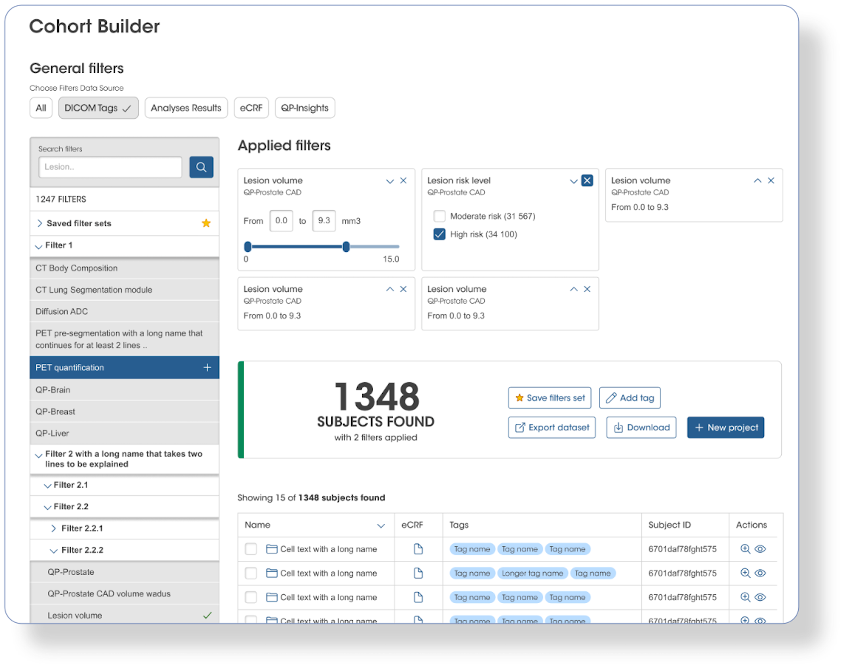
A precision search engine that unifies clinical variables, DICOM metadata, and all previously computed analyses, enabling optimized cohort identification before a project begins.

eCRF interoperability
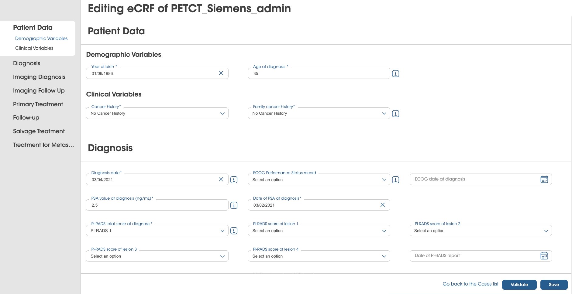
Imaging biomarker results flow into electronic data capture in real time, with an eCRF configured for your specific study that enables multi-omic model development.

Strong track record

QP-Insights® has consistently delivered dependable, high-performance solutions that address the full spectrum of our partners’ data management, storage, and processing needs—ensuring their imaging and multi-omics workflows run smoothly, securely, and at scale.
QP-Insights® is the backbone platform powering nine EU-wide research consortiums, including EUCAIM, a cornerstone of Europe’s Beating Cancer Plan, and is already trusted by major biopharmaceutical companies, academic institutions and hospitals. These collaborations support our efforts to accelerate precision medicine and patient health.
Quibim’s cloud medical platform: the AI-driven medical technology for data management and processing
As value-based care models evolve, there is a greater need to provide the right treatment for the right patient at the right time. We design end-to-end solutions working with different healthcare partners to analyze and extract new insights from multi-modality images by linking electronic health records and patient outcomes with imaging biomarkers. The assets provided improve patient stratification and generate clinical endpoints.
Imaging AI Suite
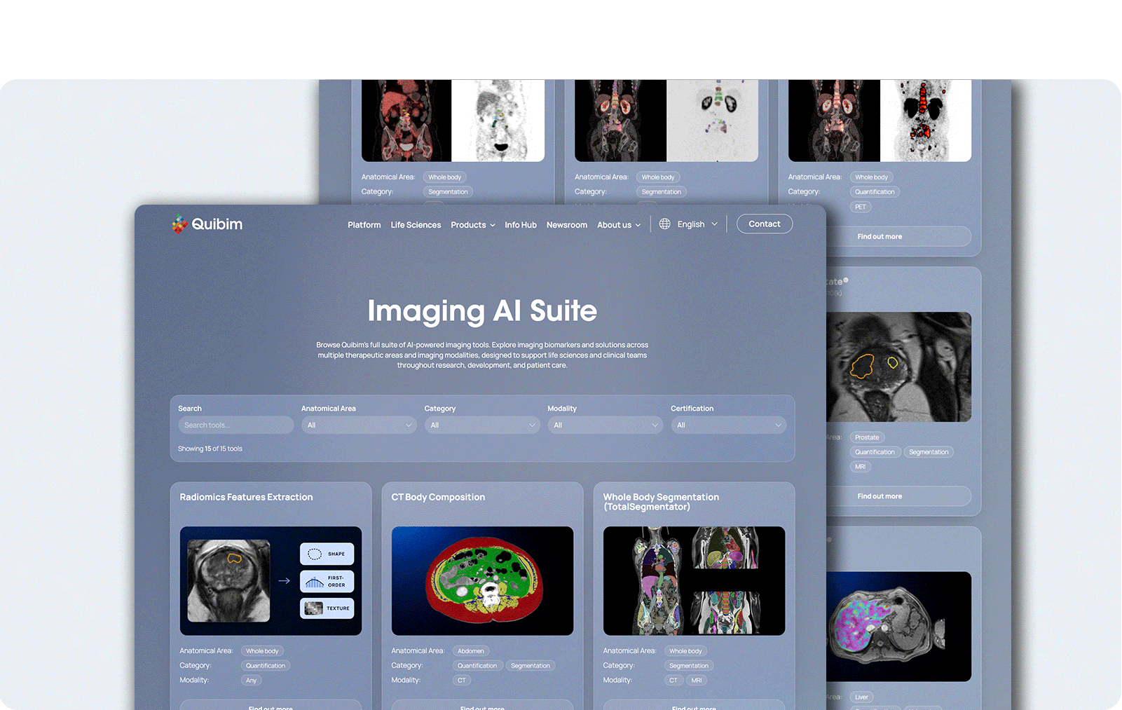
Validated tools across therapeutic areas and modalities, ready to run on any study in your project.
Radiomic features extraction
Extract shape, first-, second- and higher-order features from any region of interest, including the tissue ring around it, where signal is often missed. Full control over normalization, resampling and which features to compute.
Body composition analysis in CT scans
Automated volumetric segmentation of muscle and intramuscular, visceral and subcutaneous fat across L2–L4, returning both volumetric and L3-level metrics.
FDG and PSMA PET whole-body segmentation
Automatic volumetric segmentation of PET-avid lesions in whole-body PSMA PET (Ga-68 and F-18) and 18F-FDG PET, with quantitative metrics per lesion. Supervise and correct in the QP-Insights viewer — built to accelerate the annotation workload of nuclear medicine readers.
Regulatory-cleared products
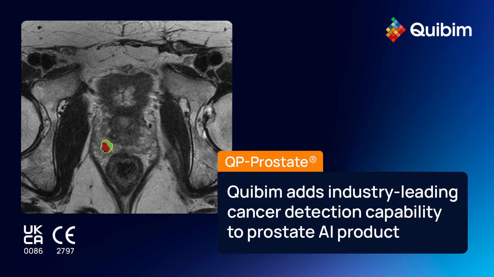
Regulatory-cleared SaMD products, viewable in the viewer, such as QP-Prostate® and QP-Breast®.
Streamlining RECIST evaluation with AI
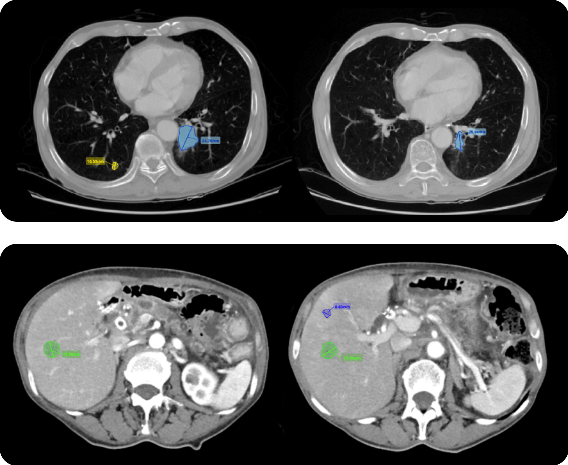
Deep learning detects, segments, measures and tracks lesions in the lungs and liver — identifying new lesions and following existing ones across every follow-up after baseline.
Radiologists keep full control: every AI measurement can be adjusted, and lesions in other anatomical regions can be added by hand.
Everything is stored in a standardized digital format as it’s produced, so the RECIST 1.1 response is calculated automatically and the final assessment report writes itself.
QP-Insights® Connector — Ask your imaging platform a question
QP-Insights® Connector links your deployment to Claude and other AI assistants over the Model Context Protocol, the open standard for connecting assistants to real systems. No export, no new interface, no ticket to the data team.
What can you ask?
- “How many subjects does PROJECT-204 have, and how do they split across sites?”
- “Which exams from my site are still waiting on analysis?”
- “Has the analysis on this study finished — and what did it measure?”
- “Tag every series in this study as QC pass.”
It sees exactly what you see. Nothing more.
- It signs in as you. Per-user OAuth 2.1 through your own QP-Insights account — the assistant inherits your QP-Insights permissions exactly.
- Nothing is stored. The connector holds no platform data and no patient data.
- Your deployment, your instance. One connector per QP-Insights environment, running inside your own infrastructure boundary — not a shared service with a queue of other people’s data in it.
Next-generation AI – More evidence per patient
Deep learning was built for a million images. A Phase II cohort has forty patients. Every research line below tackles that gap from a different angle — using the data you already hold, generating the cases you will never recruit, and transferring enough learnings from our studies that yours don’t have to start from zero.
Automated AI model builder
The imaging data collected for a primary endpoint already contains quantitative signals about that patients that can say who responded, who progressed, and who should never have been enrolled, often months before conventional endpoints. A stratification signature that identifies likely responders before randomization is the difference between a 700-patient trial and a 400-patient one, and between a Phase II that reads out ambiguously and one that reads decisively.
Leveraging the Model Builder at a cohort you already manage within the platform you can bring imaging biomarkers, radiomics features and clinical variables together in a single harmonized table. Configure the experiment once, let the search run unattended, and automatically compare models on a ranked leaderboard with per-model explainability. Your scientists drive this themselves end to end and, all data, models and results remain fully inside the environment.
Synthetic imaging data
Imaging endpoints usually fail for two reasons: too few patients with some specific characteristics, and models that perform beautifully at one site but falls over when exposed to different set of patients at a different site.
Generative models produce studies for presentations that are rare in any single cohort — uncommon mutations, atypical morphology — so a program in a small indication is no longer disqualified from an imaging endpoint simply because of the size of its own population.
Imaging foundation models
The reason exploratory imaging endpoints get cut from protocols is not due to their scientific value but because they require annotation. A new biomarker conventionally means assembling a dedicated cohort and paying radiologists to label thousands of studies. That requires a timeline and a budget few protocols rarely survives during review, so the question gets cut, and the scans get used once.
A base model trained across modalities and anatomical areas changes that arithmetic. Instead of thousands of labels, adapting it to a new question takes a few hundred annotated studies, which brings a new endpoint inside the window of the trial rather than after it. At Phase I or II scale, where conventional deep learning simply does not have the data to work with, foundation models are the difference between an imaging endpoint and having none.
And it works portfolio-wide. Being modality- and organ-agnostic, one model serves programs across indications, including a novel clinical indication with no pre-existing model behind it.