
Imaging AI Suite
Browse Quibim's full suite of AI-powered imaging tools. Explore imaging biomarkers and solutions across multiple therapeutic areas and imaging modalities, designed to support life sciences and clinical teams throughout research, development, and patient care.
Search
Anatomical Area
Category
Modality
Certification
Radiomics Features Extraction
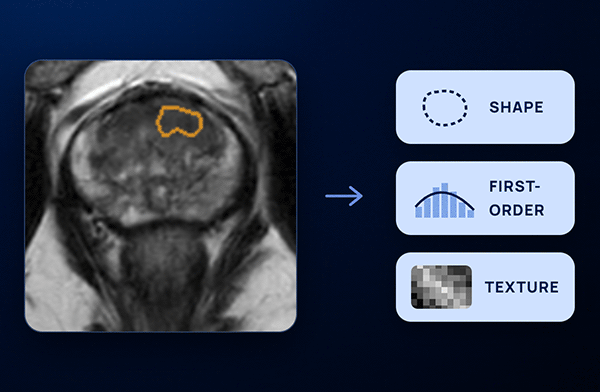
Description: Quantification of radiomics features (shape, first order, second order, and higher order features) given a pre-defined region of interest (ROI). Furthermore, additional insights from the ROIs' peripheral ring can be obtained by quantifying radiomics features in the tissue periphery. This tool provides the user with full control of the analysis through input parameters to activate/deactivate image normalization, apply resizing to isotropic voxel, force 2D analysis, or decide which radiomics features to compute.
CT Body Composition
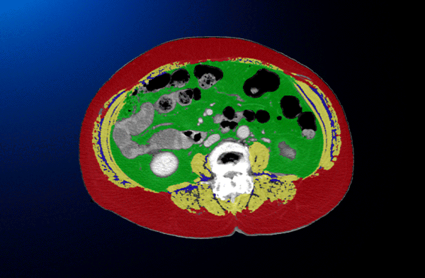
Description: AI-based evaluation of body composition in CT scans. It automatically performs the volumetric segmentation of muscle, and intramuscular, visceral, and subcutaneous adipose tissue in the abdominal area (L2-L4), providing quantitative values with volumetric and L3-level metrics.
Whole Body Segmentation (TotalSegmentator)
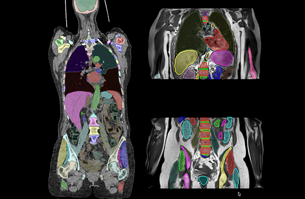
Whole Body Segmentation (TotalSegmentator)
Description: Based on the well-known TotalSegmentator solution this tool can be used for the segmentation of most major anatomical structures in CT (117 structures) and MRI (50 structures).
Scientific Publications:
https://pubs.rsna.org/doi/10.1148/ryai.230024
FDG-PET Segmentation
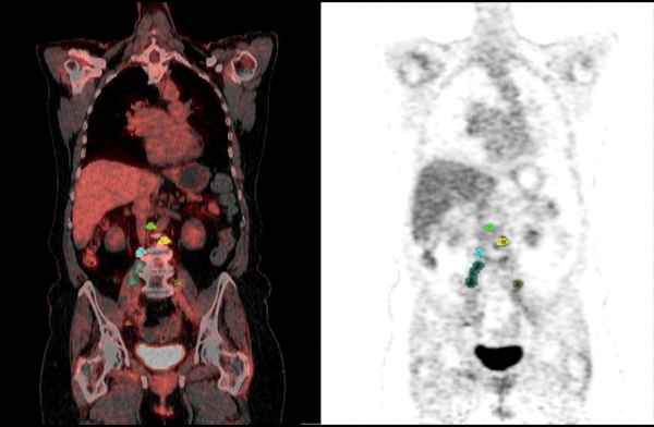
Description: Automatic segmentation of tumoral lesions in whole-body 18F-FDG PET scans. The tool automatically performs the volumetric segmentation of PET-avid lesions. PET-derived quantitative metrics are extracted from each detected lesion. This solution can be used to help nuclear medicine doctors during the annotation process using our QP-Insights viewer.
PSMA-PET Segmentation
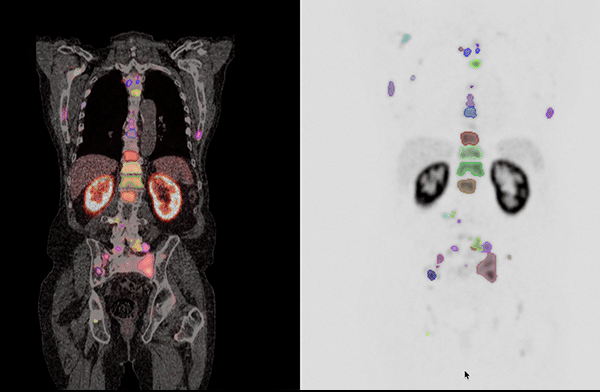
Description: Automatic segmentation of tumoral lesions in whole-body PSMA PET scans (Ga-68 and F-18). The tool automatically performs the volumetric segmentation of PET-avid lesions. QP-Insights viewer can be used for supervision. PET-derived quantitative metrics are extracted from each detected lesion. This solution can be used to help nuclear medicine doctors during the annotation process using our QP-Insights viewer.
PET Quantification
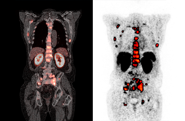
Description: Quantification of radiotracer-agnostic PET features (SUVmax, SUVmean, SUVpeak, Metabolic Tumor Volume, PSMA Tumor Volume, Total Lesion Glycolysis, Total Lesion Uptake, among others) given an input segmentation mask. Features are extracted per ROI. Normalised values to a reference tissue can also be computed together with additional insights from the ROIs' peripheral ring, quantifying the tissue periphery.
CT Lung Lesion Segmentation and Tracking
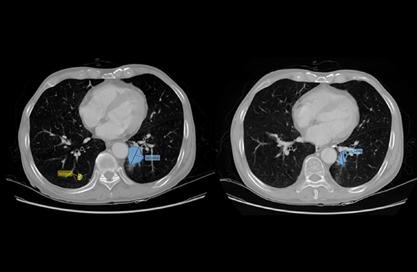
Description: Automatic segmentation, measurement, and tracking of lung lesions in CT scans. The tool automatically segments both primary tumors and metastases as 3D volumes and quantifies their short and long axis. In follow-up scans, it automatically tracks all lesions to measure any changes in volume and size. This solution can be used to help radiologists during the annotation process using our QP-Insights viewer.
Scientific Publications:
https://doi.org/10.1186/s41747-025-00617-7
,
https://ieeexplore.ieee.org/document/11515463
CT Liver Metastasis Segmentation and Tracking
CT Liver Metastasis Segmentation and Tracking
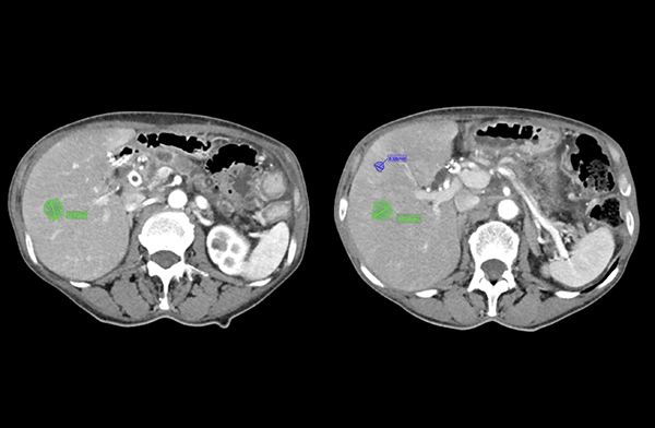
Description: Automatic segmentation, measurement, and tracking of liver metastasis in CT scans. The tool automatically segments tumor 3D volumes and quantifies their short and long axis. In follow-up scans, it automatically tracks all lesions to measure any changes in volume and size. This solution can be used to help radiologists during the annotation process using our QP-Insights viewer.
Scientific Publications:
https://ieeexplore.ieee.org/document/11515463
QP-Prostate®
CE / UKCA / 510(k)
QP-Prostate®
CE / UKCA / 510(k)
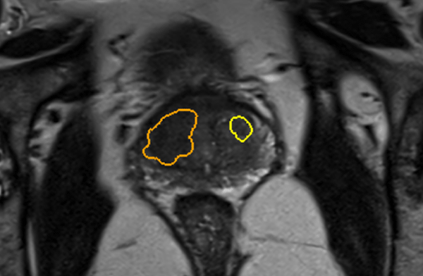
Description: Automatic segmentation of prostate gland and clinically significant prostate cancer lesions.
More info:
https://www.quibim.ai/qp-prostate
QP-Breast®
CE / UKCA
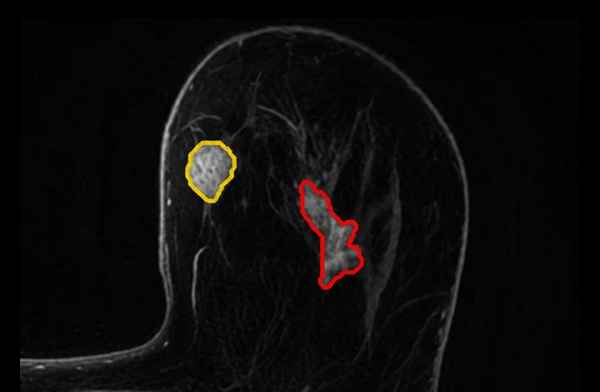
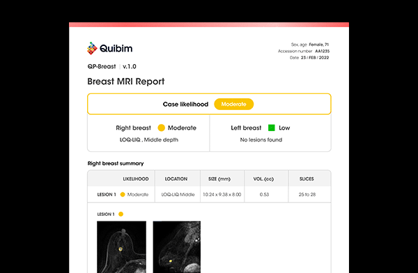
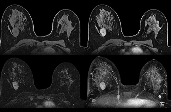
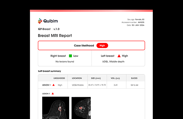
Description: AI-powered solution designed to support radiologists in breast MRI interpretation.
More info:
https://quibim.ai/qp-breast/
QP-Brain®
CE / UKCA / 510(k)
QP-Brain®
CE / UKCA / 510(k)
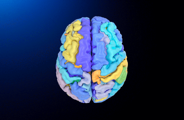
Description: Automatic segmentation and volumetric quantification of brain structures and white matter hyperintensities.
More info:
https://quibim.ai/qp-brain/
QP-Liver®
CE / UKCA
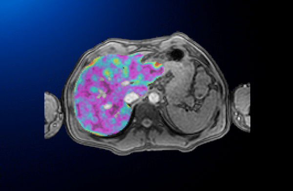
Description: Automated quantification of liver fat fraction and iron content.
More info:
https://quibim.ai/qp-liver/
Diffusion ADC
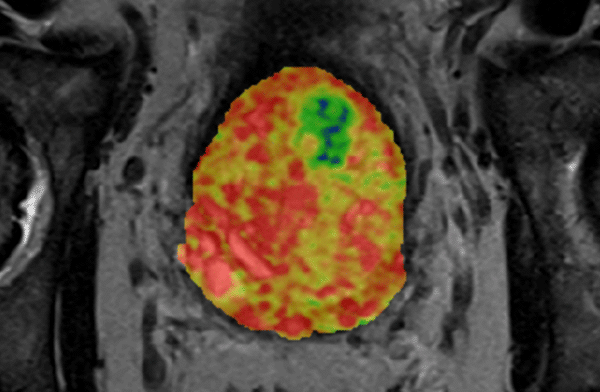
Description: Quantification of Apparent Diffusion Coefficient (ADC) on Diffusion Weighted MR Image (DWI). The tool outputs the volumetric ADC parametric map along with ROI-based measurements. Additional insights from the ROIs' peripheral ring can be obtained by quantifying ADC values in the tissue periphery.
Lung Densities
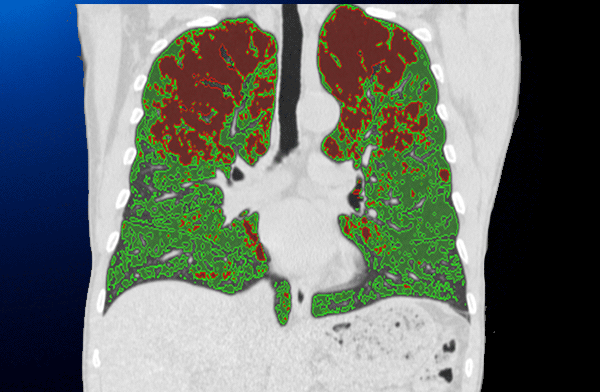
Description: Automatic quantification of lung emphysema and low densities in thoracic CT scans. Two strategies are followed: first, a fixed threshold is applied; then, our patented adaptive threshold calculation solution is used for quantification, reducing any bias introduced by the scanner or the patient characteristics. Per-lobe quantitative volumes and percentages are computed.
Bone Microarchitecture Quantification
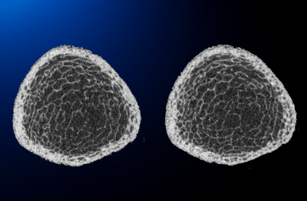
Bone Microarchitecture Quantification
Description: Automatic quantification of trabecular bone microarchitecture. Imaging features such as the bone volume to total volume ratio, trabecula thickness, pore size, trabecula number, or the fractal dimension are quantified for bone quality assessment. The processing pipeline has been adapted to support MR, CT, or X-ray images.